
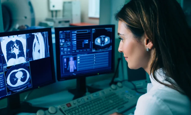
Medical imaging has always been a cornerstone of modern healthcare, but the way we capture and interpret those images is changing dramatically. From the days of grainy black-and-white X-rays to today’s crisp 3D scans, the evolution has been relentless. Now, Diag Image is leading a new wave of transformation one that doesn’t just make images clearer but makes them smarter.
This technology represents the fusion of high-resolution imaging with artificial intelligence and clinical workflow integration, turning what was once a static picture into an interactive, data-rich diagnostic tool. In the fast-paced world of medicine, where time can mean the difference between recovery and decline, Diag Image is helping clinicians make faster, more informed decisions.
Table of contents
- Why Diag Image is a Game Changer in Healthcare
- The Core Technologies Behind Diag Image
- Real-World Case Studies: Diag Image in Action
- How Diag Image Improves Patient Outcomes
- Applications Across Specialties
- The AI Advantage: Learning with Every Scan
- Ethical Considerations and Safeguards
- Looking Ahead: The Future of Diag Image
- Conclusion
- FAQs
Why Diag Image is a Game Changer in Healthcare
In traditional settings, a patient might undergo a scan, wait hours or even days for a radiologist’s review, and only then receive a treatment plan. This gap between image capture and diagnosis can be critical, especially in emergency medicine.
Diag Image changes this model by integrating real-time image processing and AI-powered analysis directly into the imaging workflow. By doing so, it delivers immediate, actionable insights a leap forward for patient care.
More than a technology upgrade, Diag Image represents a philosophical shift: from “take the picture and wait” to “take the picture and act.”
The Core Technologies Behind Diag Image
The power of Diag Image lies in its synergy between imaging hardware and computational intelligence. While familiar modalities like MRI, CT, ultrasound, and X-ray remain the foundation, Diag Image enhances them with several advanced capabilities:
AI-Driven Image Analysis
At the heart of Diag Image is an AI engine trained on millions of annotated medical images. It doesn’t just detect anomalies it can compare current scans to historical datasets, spotting subtle changes over time that may escape the human eye.
High-Resolution, Low-Latency Imaging
Patients benefit from sharper images without longer scan times. For those in pain or distress, reduced scan duration is more than a convenience it’s a necessity.
Real-Time Reporting
By embedding AI analysis within the imaging device it can generate preliminary reports within minutes, giving physicians a head start on diagnosis and treatment.
EHR Integration
Diag Image seamlessly syncs with electronic health record systems, ensuring that imaging results are immediately accessible alongside other patient data.
Real-World Case Studies: Diag Image in Action
Adding real-world examples highlights how Diag Image is not just theory but practice.
Case Study 1: Stroke Detection in Minutes
Traditionally, his CT scan would be reviewed manually, a process taking 30–60 minutes. With, AI algorithms flagged a clot in the middle cerebral artery within three minutes of image acquisition. The stroke team was activated immediately, and the patient received clot-dissolving therapy within the critical “golden hour.” Follow-up showed minimal neurological damage a testament to the life-saving power of rapid diagnosis.
Case Study 2: Rural Oncology Screening
A rural clinic without an on-site radiologist adopted Diag Image’s cloud-based solution. A patient’s mammogram revealed a suspicious lesion, instantly flagged by the AI. The image and findings were transmitted to a specialist 200 miles away, who confirmed the diagnosis. Early detection allowed for a lumpectomy rather than a full mastectomy, significantly improving the patient’s quality of life.
Case Study 3: Orthopedic Surgery Planning
An orthopedic center used Diag Image to scan a complex knee injury. The AI’s 3D reconstruction allowed the surgeon to plan a minimally invasive procedure, reducing recovery time from six months to three. The patient returned to competitive sports — something not initially thought possible.
How Diag Image Improves Patient Outcomes
Beyond speed and clarity it directly impacts treatment success rates. Its ability to triage cases ensures that life-threatening conditions are prioritized. By minimizing false positives, it reduces unnecessary procedures. And its detailed imaging helps specialists choose the least invasive, most effective treatment paths.
For hospitals, this means lower operational costs and better patient satisfaction. For patients, it means faster answers, fewer uncertainties, and better health outcomes.
Applications Across Specialties
Diag Image is not a one-size-fits-all tool it adapts to various medical fields:
Oncology
Detects tumors at sub-millimeter levels, enabling earlier intervention and personalized treatment planning.
Cardiology
Improves accuracy in detecting arterial blockages and heart defects, guiding both emergency interventions and long-term care.
Neurology
Tracks the progression of neurodegenerative diseases, ensuring treatment adjustments are made proactively.
Orthopedics
Provides high-resolution visualization of bones and soft tissue for precise surgical planning.
The AI Advantage: Learning with Every Scan
One of Diag Image’s strengths is its machine learning foundation. Every scan processed contributes to its growing dataset, making its predictions more accurate over time.
For example, in lung imaging it can distinguish between benign and malignant nodules with increasing accuracy. This not only improves patient care but also builds clinician trust in AI-assisted diagnostics.
Ethical Considerations and Safeguards
Introducing AI into healthcare comes with responsibilities. Diag Image follows a human-in-the-loop model, ensuring no AI-generated finding is finalized without expert review.
The system also prioritizes explainable AI, showing exactly why an anomaly was flagged, which supports transparency and accountability. On the privacy front, all data is encrypted and anonymized, meeting HIPAA and GDPR compliance.
Looking Ahead: The Future of Diag Image
The future holds exciting possibilities:
- Predictive analytics that anticipate disease progression
- Integration with wearable devices for continuous monitoring
- Expansion into underserved regions through cloud-based imaging
As these capabilities mature it could become a central hub of diagnostic intelligence, serving not just hospitals but entire healthcare networks.
Also Read What Is Gastroshiza? Causes, Diagnosis & Care
Conclusion
Diag Image is more than just an imaging system it’s a catalyst for a new era of diagnostics. By combining precision imaging, AI-driven analysis, and real-world clinical integration, it is helping healthcare providers deliver faster, more accurate, and more compassionate care.
In a world where seconds can save lives, technologies like aren’t just innovations — they are necessities. As it continues to evolve, we can expect its impact to extend far beyond radiology, shaping the very way we practice medicine.
FAQs
1. What is Diag Image’s main purpose?
Diag Image enhances medical imaging with AI-powered analysis for faster and more accurate diagnostics.
2. Can Diag Image be used in small clinics?
Yes, its cloud-based version is ideal for clinics without in-house radiologists, making advanced diagnostics accessible in remote areas.
3. How accurate is Diag Image compared to traditional methods?
Studies show it can match or exceed the accuracy of human radiologists, especially in early disease detection, though it always works alongside human experts.
4. Does Diag Image replace doctors?
No. It supports clinicians by providing faster insights, but final decisions remain with trained medical professionals.
5. Is patient data safe with Diag Image?
Yes, it follows strict HIPAA and GDPR compliance, with full encryption and anonymization protocols.
6. What medical fields benefit most from Diag Image?
Oncology, cardiology, neurology, orthopedics, and emergency medicine all see significant improvements from its use.